
Dr David Bhella (CVR programme leader) and Dr Ed Hutchinson (MRC research fellow at the CVR) tell us about influenza and its filamentous nature.
Even after eighty years of studying them, we still tend to forget what influenza viruses look like. In a paper published this week, the Hutchinson and Bhella labs (together with collaborators at the University of Oxford) draw attention to a frequently overlooked feature of influenza viruses: their tendency to form extremely long, thin virus particles.
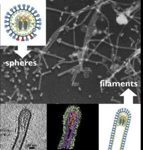
Although some other viruses are well-known for forming long filaments – Ebola viruses, for example – influenza viruses are best known for forcing their hosts to produce virus particles shaped like tiny spheres or kidney beans. These small round particles form readily in the lab, and have been the focus of the vast majority of influenza research. But just because something is easy to observe in the lab, it doesn’t mean that it tells the whole story about actual infections.
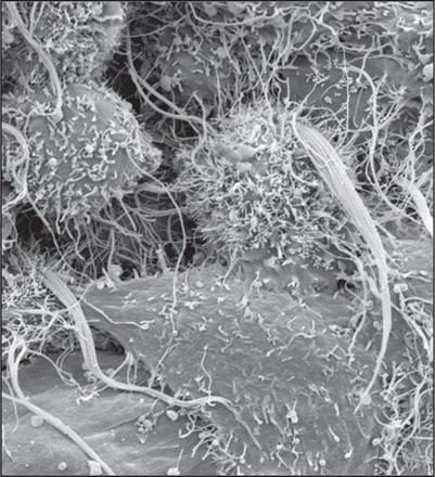
Since the 1940s, occasional reports have noted that clinical isolates of influenza contain both spherical particles and extremely long filaments, some of them hundreds of times longer than the spheres (Figure 1). The result is that when you’re infected with influenza, cells in your throat begin to sprout dense clumps of hair-like viruses (Figure 2). The ability to make these filaments is typically lost as influenza viruses evolve to grow in laboratories rather than in throats, and so until recently they have been little more than a virological footnote.
Recent work is changing this. Studies in animals have shown that filaments do give the virus an advantage in respiratory infections, and structural studies, including recent work by the Bhella group have shown that filaments have a characteristic architecture. By looking in more detail at the structure and composition of these filaments, the Hutchinson and Bhella labs now hope to start linking these remarkably extended forms to a function. Why it benefits influenza to elongate some of its virus particles until they are hundreds of times longer than others is currently a mystery. But, as this literature review highlights, we now know enough about the basic properties of filaments to give that mystery some serious thought.
**************************************************
The MRC – University of Glasgow Centre for Virus Research has a range of research groups studying influenza – the structural biology of filamentous influenza particles (David Bhella and Ed Hutchinson), understanding the molecular and evolutionary mechanisms of influenza emergence into new hosts (Pablo Murcia), and how influenza interacts with the host cell during infection (Chris Boutell).